Describe Patient Preparation for Radiographic Exposures
Assessment and Preparation 1. Identify safety measures to reduce operator x-radiation exposure.

Take A Bite Out Of Dental Radiology Positioning For Picture Perfect Views Today S Veterinary Nurse
You want to make sure that they have any jewelry removed that could interfere with the radiographs goes for all.

. Restricting the area of the patient irradiated is one of the most effective ways to reduce patient exposure. HOBBS MSRS RTRCTMR After completing this article the reader should be able to. Adjust the headrest to support the patients head.
Describe patient preparation for radiographic exposures eg inspect the patients head and neck for removable appliances and foreign objects. It is important for the radiographer to determine the amount of mAs needed to produce a diagnostic image. Have patient bite down on cotton rollbite block Make sure patient is standing straight up with legs shoulder width apart Make sure patients head is straight up eyes parallel to floor when looking straight Have patient relax shoulders Have patient close mouth relax lips b.
50 mA 200 ms 02 s 10 mAs. Describe patient preparation for radiographic exposures Describe the use and purpose of intraoral and extraoral radiographic images Select an appropriate radiographic survey to examine or view conditions teeth or landmarks Describe technique. Remove dentures tongue pricing missing teeth is isolated earrings.
Position the patient upright in the chair. Describe patient preparation for radiographic exposures Describe use and purpose of intraoral and extraoral radiographic images Select appropriate radiographic survey to examine or view conditions teeth or landmarks Describe technique modifications based on anatomical variations and clinical conditions Select equipment for radiographic technique Describe how to acquire. Two patient identifiers 15.
Exposure radiographic positioning and positioning terminology and patient care and management. Radiation Health and Safety in Dentistry is a comprehensive course that explains patient and operator preparation and safety during radiographic exposures and provides the knowledge necessary to identify best radiographic technique based on patients diagnostic needs. Radiographers must understand the rationale behind the examination.
Describe patient preparation for radiographic exposures Describe use and purpose of intraoral and extraoral radiographic images Select appropriate radiographic survey to examine or view conditions teeth or landmarks Describe technique modifications based on anatomical variations. Recognize the precautions to be. CSLO 3 Demonstrate an understanding of the patient preparation required prior to performing each.
200 mA 50 ms 005 s 10 mAs. Adjusting Milliamperage and Exposure Time to Maintain mAs. Apply universal precautions with every patient 14.
Apply critical thinking skills in performing the patient assessment and patient care. Describe the anatomical relationships of various organs in the chest. To floorAla-tragus line parallel to floor.
Measuring Dental Assisting Excellence Radiation Health and Safety RHS Exam Blueprint I. There should be at least three edges of collimation on each film. Otherwise radiographs of diagnostic value cannot be produced.
To maintain the mAs use. Explain the patient preparation for routine radiographic examinations 17. Describe use and purpose of intraoral and extraoral radiographic images including but not limited to.
Any individual holding or supporting a person during radiation exposure should wear protective gloves and apron with a minimum of 025 millimeters lead equivalent. Adjust the chair to comfortable working height for operator. The preparation depend on the type of ultrasound procedure Some preparations include drinking a quart of water before the test to obtain better images.
Tap card to see definition. You should expose as little of the patient as possible and evidence of use of the collimator should be present on your finished radiographs. Following this course students will be able to.
Define spell and pronounce the terms listed in the vocabulary. Click again to see term. Describe patient care considerations for the patient with a chest tube and water-sealed drainage.
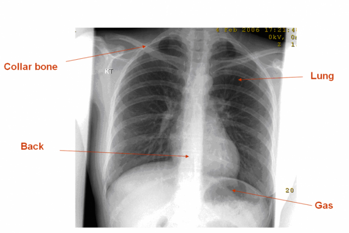
Describe exposure technique ie patient positioning. Describe the cassette and film image receptor system and explain its function in radiography. Identify the basic anatomy seen on a chest radiograph.
100 mA 100 ms 01 s 10 mAs. Describe Patient Preparation for radiographic exposures. Identify methods and barriers of communication and describe how each may be used or overcome effectively during patient education 16.
In addition the. EXPOSE AND EVALUATE A. Chest Radiography for Radiologic Technologists DAN L.
Remove all objects from the patients mouth that may interfere with image exposure including eyeglasses. Address patient concerns about radiation including informed consent or patient refusal of radiography Identify sources of x-radiation to operatorsother staff while exposing image receptors. Effective 01012015 4203 Dental Assisting National Board Inc.
For a biopsy do not eat or drink anything past midnight the night before the exam. The patient must be positioned and the exposure factors selected according to the region involved and the radiographic characteristics of the existent abnormality. Describe patient preparation for radiographic exposures eg inspect the patients head and neck for removable appliances and foreign objects.
Describe the basic positioning requirements for a chest exam. Healthcare personnel assisting with radiological procedures must avoid holding the patient manually during a radiographic study due to the risk of direct beam exposure. Click card to see definition.
Identify the principal components of radiographic equipment. Radiation Health and Safety EXAM BLUEPRINTS AND SUGGESTED REFERENCES Page 2 2015 Dental Assisting National Board Inc.

1100 Mcq In Dentistry With Answers Dentistry Panoramic Radiograph Mcq

3677 Pdfs Review Articles In X Ray Radiography

Chest X Ray Chest Radiography Nursing Responsibilities Nurseslabs

Endodontic Radiology Pocket Dentistry

Portable Radiography Radiology Reference Article Radiopaedia Org
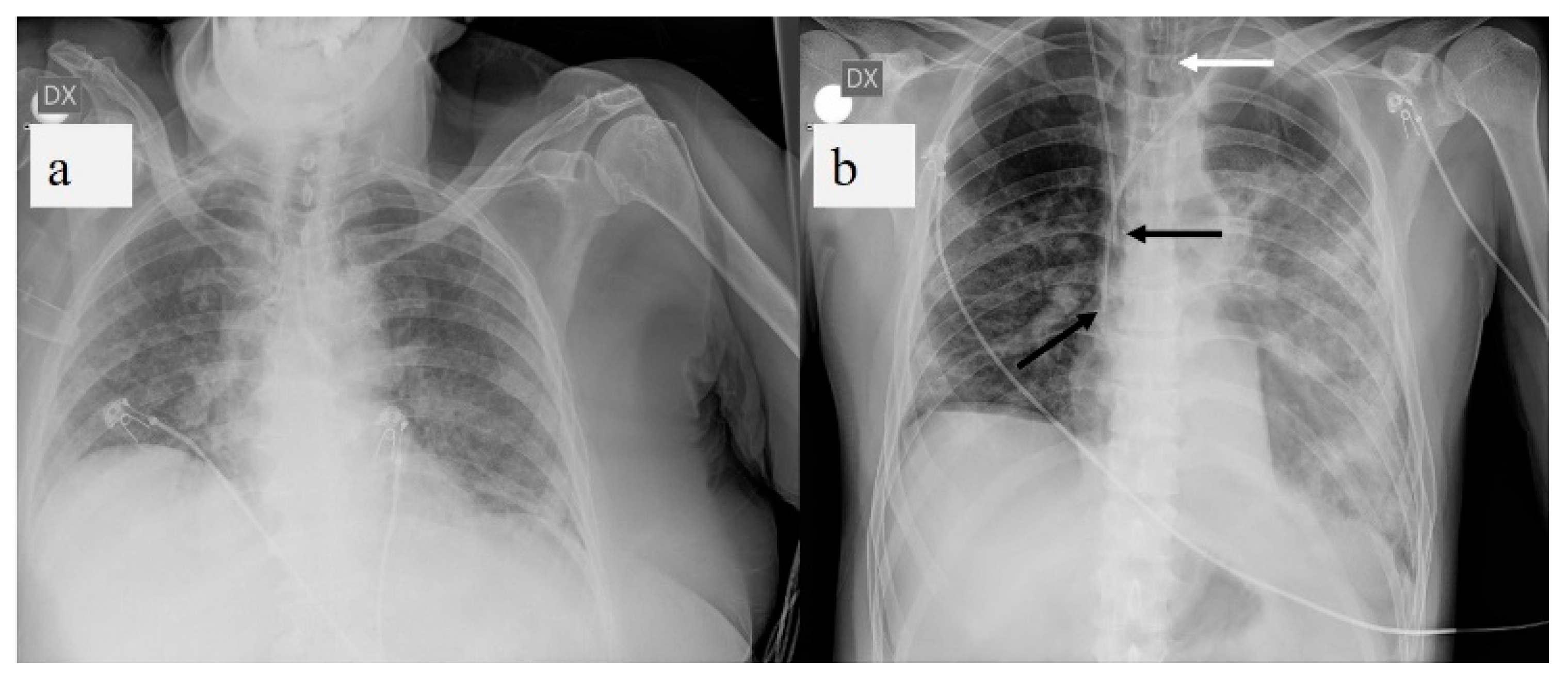
Characterisation Of Hemidiaphragm Dysfunction Using Dynamic Chest Radiography A Pilot Study European Respiratory Society
1 Plain Radiographic Imaging Radiology Key
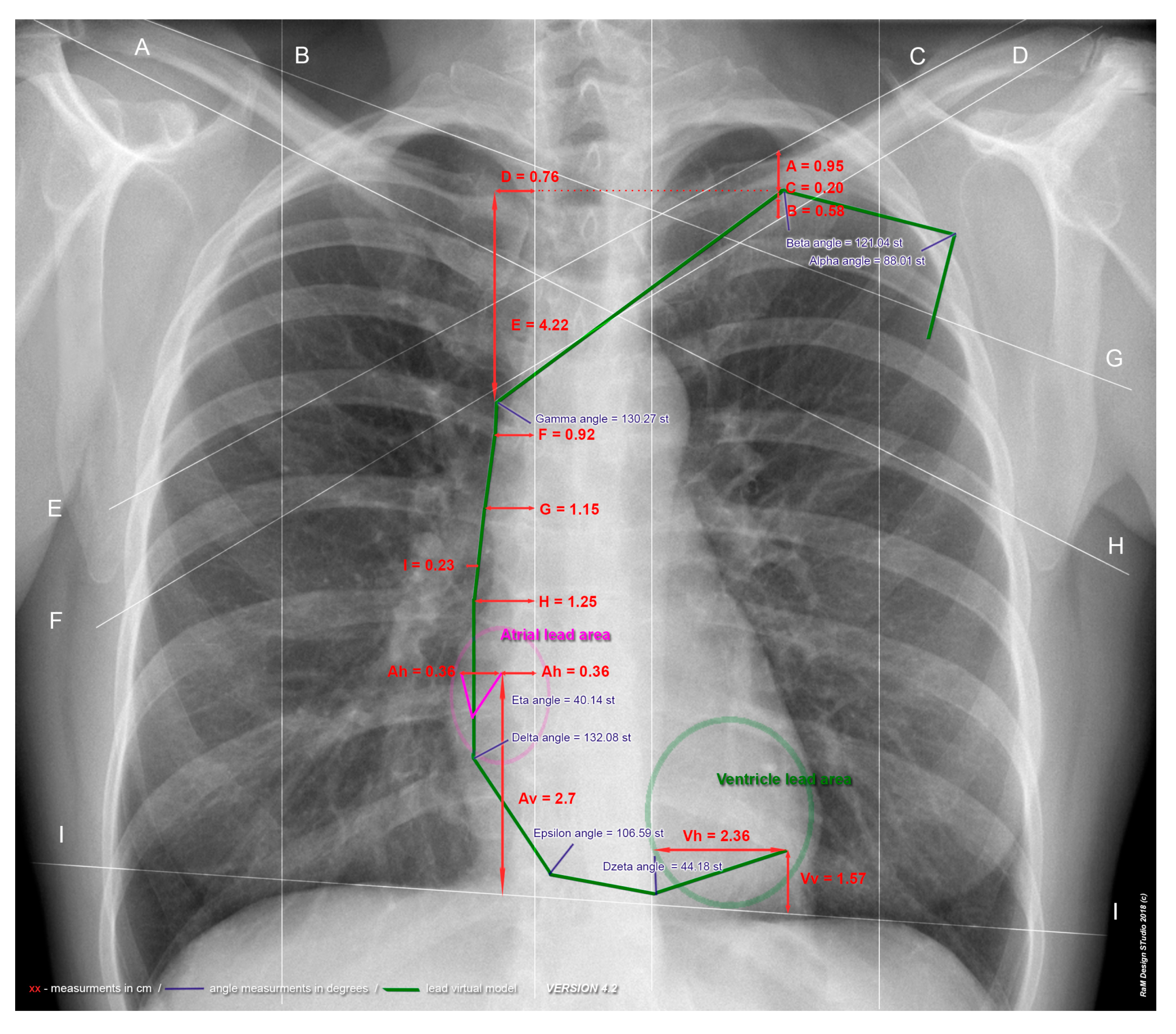
Medicina Free Full Text New Method Of Cardiac Lead Evaluation Using Chest Radiography Html
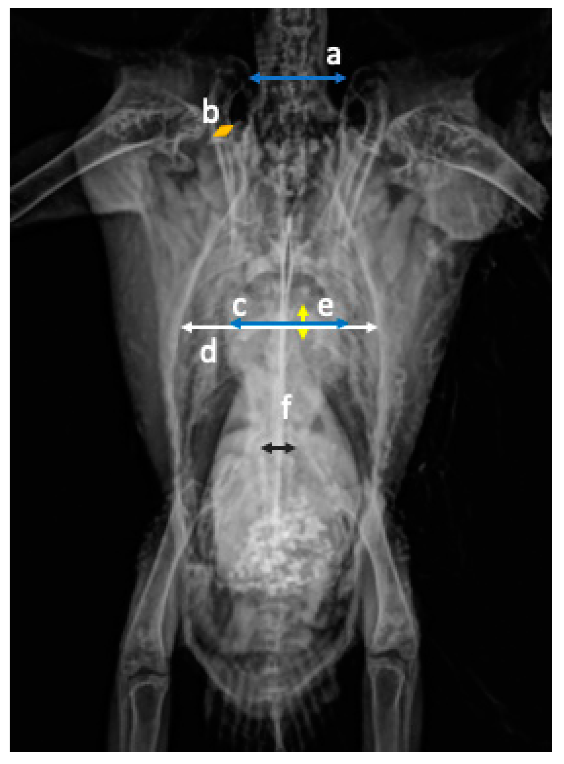
Animals Free Full Text Radiographic Measurements Of The Cardiac Silhouette And Comparison With Other Radiographic Landmarks In Wild Galahs Eolophus Roseicapilla Html
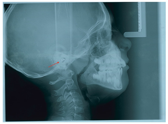
Children Free Full Text Incidental Finding In Pre Orthodontic Treatment Radiographs Of An Aural Foreign Body A Case Report Html
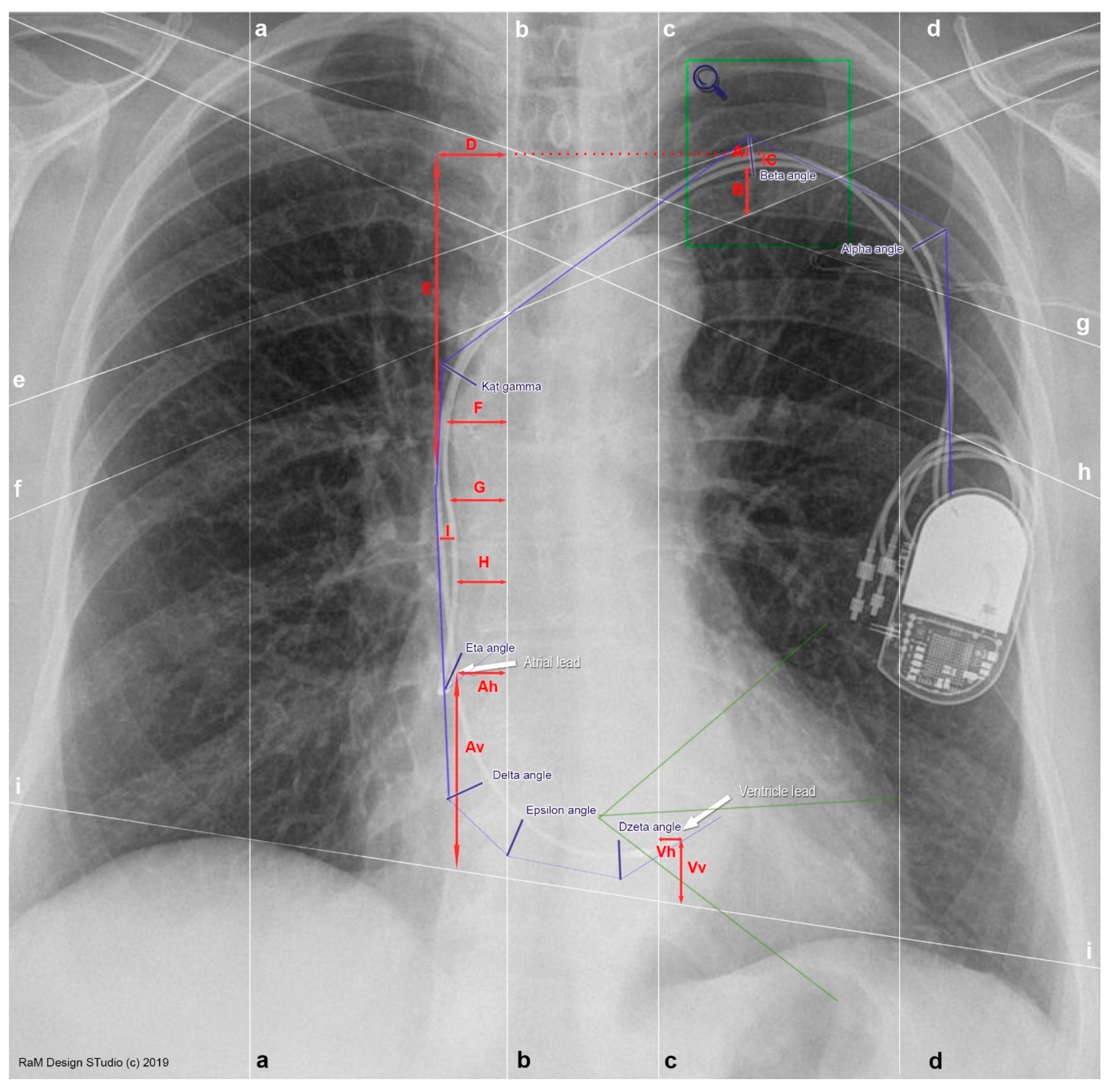
Medicina Free Full Text New Method Of Cardiac Lead Evaluation Using Chest Radiography Html
Plain Radiograph X Ray Insideradiology

Endodontic Radiology Pocket Dentistry
Patient Preparation Panoramic Radiographs Technique Anatomy Review Continuing Education Course Dentalcare Com

Pin On Radiology

Endodontic Radiology Pocket Dentistry

Panoramic Radiography And Patients With Disability A New Simple Breathing Technique To Reduce Common Airspace Error Scott Journal Of Medical Radiation Sciences Wiley Online Library
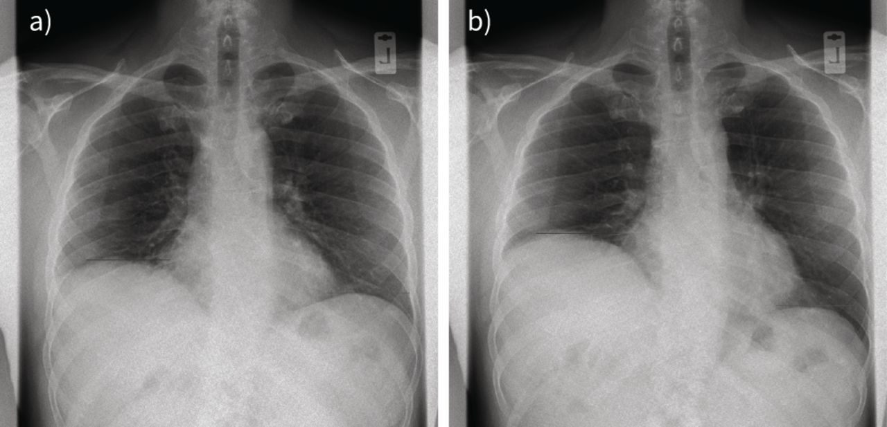
Diagnostics Free Full Text A Pictorial Review Of The Role Of Imaging In The Detection Management Histopathological Correlations And Complications Of Covid 19 Pneumonia Html

Examining Practitioners Assessments Of Perceived Aesthetic And Diagnostic Quality Of High Kvp Low Mas Pelvis Chest Skull And Hand Phantom Radiographs Journal Of Medical Imaging And Radiation Sciences


Comments
Post a Comment